


Multimodal Imaging of Carotid Plaques
To raise the bar for stroke care, it is critical that we understand the biomechanics of how and why carotid plaques rupture and cause stroke. The TNT Lab is conducting trailblazing research in multimodal carotid imaging to understand the formation of atherosclerotic plaques within vessels.
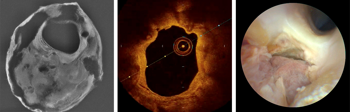
Multimodal images of carotid arteries. From left to right: microMRI, Intravascular OCT (Optical Coherence Tomography), laser angioscope.
Disruption of the inner surface of a vulnerable carotid plaque is considered the precipitating factor that culminates in the formation of clots leading to stroke. At the TNT lab, we have characterized the interplay of forces driving flow, tissue interactions, and phenotypical hallmarks resulting from plaque disruption and healing.
Based on extensive structural analysis and computational flow dynamic studies, we have shown that carotid plaque disruption is a hemodynamically driven phenomenon triggered by fibrous cap weakening, delamination, and ulceration followed by penetration of blood and thrombosis.
In this paradigm, intraplaque hemorrhage is of major importance and represents the 'smoking gun' of the root-cause of a stroke.
To ensure that this new knowledge impacts patient's lives, the lab has leveraged cutting edge preclinical imaging tools such as micro-CT, micro-MRI and scanning electron micrography to better characterize unstable plaque phenotypes.
Additionally, we are pioneering the use of next-generation intravascular imaging platforms to visualize at unprecedented resolution these structural changes at the lumen-wall interface of vulnerable and disrupted plaques. In particular, the lab is investigating the use of multimodal carotid imaging, such as laser scanning fiber endoscopy and optical coherence tomography (OCT).
Furthermore, our group has used and validated advanced MR sequences to non-invasively detect carotid intraplaque hemorrhage in patients with stroke and is pushing forward the first clinical trial to implement this approach.
Our translational work is fueled by national and international collaborations including the Carotid Registry of Imaging and Pathology (CRIP), which was started and is run by the TNT Lab. This multicenter collaborative biobank receives carotid endarterectomy specimens from several major academic U.S.-based institutions and conducts pioneering translational research in carotid artery disease and vascular biology.
To translate these new disease insights and technological advancements into evidence-based medicine, the lab is working with National Institutes of Health (NIH) to launch the SYmptomatic Non-stenotic Carotids with High-Risk imaging biOmarkers (SYNCHRO) trial.
This is a StrokeNet National Institute of Neurological Disorders (NINDS)-supported trial that challenges the dogma of carotid stenosis by using modern biomarkers of plaque vulnerability to risk-stratify people with strokes. This multicenter, hypothesis-driven imaging biomarker study will evaluate the rate of recurrent stroke in patients with symptomatic nonstenotic carotid (SyNC) managed with intensive medical therapy.
This trial aims to provide the largely needed evidence that aligns the clinical management of people with carotid artery disease with contemporary understanding of plaque disruption and thrombo-embolization. Successful completion of this trial will lead to a paradigm shift in the management of carotid artery disease to prevent strokes, disability and death.
Savastano LE, Seibel E. Scanning fiber angioscopy: A multimodal intravascular imaging platform for carotid atherosclerosis. https://academic.oup.com/neurosurgery/article/64/CN_suppl_1/188/4093202 Neurosurgery. 2017; doi:10.1093/neuros/nyx322.
Savastano LE, Zhou Q, Smith A, Vega K, Murga-Zamalloa C, Gordon D, McHugh J, Zhao L, Wang M, Pandey A, Thompson BG, Zu J, Zhang J, Chen, YE, Seibel, EJ, Wang TD. Multimodal laser-based angioscopy for structural, chemical and biological imaging of atherosclerosis. https://www.nature.com/articles/s41551-016-0023 Nature Biomedical Engineering. 2017; doi:10.1038/s41551-016-0023.
Savastano LE, Chaudhary N, Murga-Zamalloa C, Wang M, Wang T., Thompson BG. Diagnostic and interventional optical angioscopy in ex vivo carotid arteries. https://academic.oup.com/ons/article/13/1/36/2734426 Operative Neurosurgery. 2017; doi:10.1093/ons/opw002.
Larson A, Nasr DM, Rizvi A, Alzuabi M, Seyedsaadat SM, Lanzino G, Huston J, Lehman VT, Savastano LE, Brinjikji W. Embolic stroke of undetermined source: The association with carotid intraplaque hemorrhage. https://www.sciencedirect.com/science/article/pii/S1936878X20307245?via=ihub JACC: Cardiovascular Imaging. 2021; doi:10.1016/j.jcmg.2020.08.007.
Larson A, Brinjikji W, Savastano LE, Huston J, Benson JC. Carotid intraplaque hemorrhage is associated with cardiovascular risk factors. https://www.karger.com/Article/FullText/508733 Cerebrovascular Diseases. 2020; doi:10.1159/000508733.
Larson A, Nardi V, Lanzino G, Brinjikji W, Scharf E, Savastano LE. Symptomatic mild carotid artery senosis. https://journals.lww.com/contempneurosurg/Fulltext/2020/09300/Symptomatic_Mild_Carotid_Artery_Stenosis.1.aspx Contemporary Neurosurgery. 2020; 10.1097/01.CNE.0000723688.52449.d5.

